
Echocardiography
- Home
- /
- Services Details
Overview
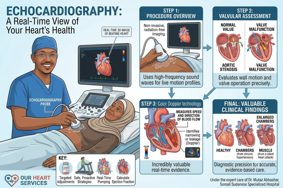
Echocardiography is a highly advanced, non-invasive imaging method that uses high-frequency sound waves to construct real-time, moving visual profiles of your beating heart. This diagnostic procedure is essential for evaluating the overall pumping efficiency of the heart muscle, calculating the ejection fraction, and observing how effectively blood moves through the cardiac chambers. It provides an immediate, highly accurate look at the physical structures of the heart without exposing the patient to radiation.
Utilizing state-of-the-art transthoracic echocardiography equipment, Dr. Abbashar meticulously examines the movement of the heart walls and the operation of the valves. This detailed structural assessment allows him to confidently identify silent valve malfunctions, congenital anomalies, or fluid accumulation around the heart muscle. By catching these subtle physical changes early, he can prescribe targeted medical adjustments or outline safe, proactive pre-surgical strategies.
During the procedure, a clear gel is applied to the chest, and a transducer probe is moved gently across the skin to transmit ultrasound waves. The returning echoes are instantly converted into detailed, live 2D and 3D images on a monitor. Dr. Abbashar uses advanced Color Doppler technology during this scan to view blood flow patterns in real time, measuring the speed and direction of blood as it passes through the heart valves to identify any leakage or narrowing.
The clinical findings from an echocardiogram are incredibly valuable. The highly detailed images allow Dr. Abbashar to assess if heart chambers have become enlarged from chronic high blood pressure, or if any portion of the heart muscle has been weakened by a silent heart attack. This diagnostic precision ensures that any subsequent treatment plan is based on highly accurate, real-time physical evidence.
Need this service?
To book a consultation about this book a visit from the button below